
Organization of Skeletal Muscle
Introduction
Skeletal muscles are specialized organs that produce voluntary body movements, maintain posture and help stabilize joints. They are attached to bones, mainly through tendons, and contract in response to signals from the nervous system.
A skeletal muscle is not simply a collection of muscle cells. It has an organized structure in which smaller components are arranged into larger units. This arrangement allows the force produced by individual muscle fibres to combine and move the skeleton efficiently.
For example, when you bend your elbow to lift a glass of water, the biceps brachii muscle contracts. Its muscle fibres generate force, which is transmitted through connective tissue and the tendon to the forearm bones.
Structural Organization of Skeletal Muscle
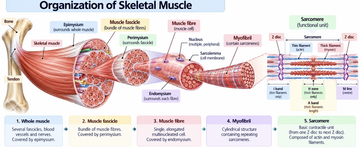
Skeletal muscle is organized into several levels, from the complete muscle down to the protein filaments responsible for contraction.
The main sequence is:
Whole muscle → Muscle fascicle → Muscle fibre → Myofibril → Sarcomere → Myofilaments
Each level has a specific structural role, and the components work together during contraction.
1. Whole Skeletal Muscle
A whole skeletal muscle is an organ made up of numerous muscle fascicles, connective tissue, blood vessels and nerves.
Each muscle is surrounded by a connective tissue covering called the epimysium. This outer covering helps maintain the muscle's shape and supports the transmission of force generated during contraction.
The muscle is commonly attached to bones through tendons, which are strong connective tissue structures that transfer muscular force to the skeleton.
Example: The biceps brachii is a skeletal muscle that helps flex the elbow and is involved in movements such as lifting an object.
2. Muscle Fascicles
A muscle fascicle is a bundle of skeletal muscle fibres within a muscle.
Each fascicle is surrounded by a connective tissue layer called the perimysium. This arrangement groups muscle fibres together and provides pathways for blood vessels and nerves.
A muscle contains multiple fascicles, and their combined activity contributes to the force generated by the whole muscle.
3. Muscle Fibres
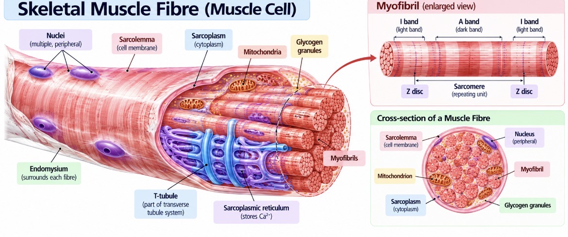
A skeletal muscle fibre is a single, elongated muscle cell. Unlike most cells, a mature skeletal muscle fibre contains multiple nuclei, usually positioned near the periphery of the cell beneath its membrane.
The cell membrane is called the sarcolemma, while the cytoplasm is called the sarcoplasm. The sarcoplasm contains myofibrils, mitochondria, glycogen and other components needed for muscle function.
Each muscle fibre is surrounded by a delicate connective tissue layer called the endomysium.
Skeletal muscle fibres appear striated under a microscope because their contractile proteins are arranged in repeating patterns.
Real-life example: When you perform repeated squats, the muscle fibres in your thigh muscles repeatedly generate force to extend your knees and hips.
4. Myofibrils
Myofibrils are long, cylindrical structures located inside muscle fibres. They contain the contractile proteins responsible for producing muscle force.
Each myofibril consists of repeating units called sarcomeres, arranged end to end. These repeating units give skeletal muscle its characteristic striped appearance.
Myofibrils contain thin and thick protein filaments arranged in a highly organized pattern.
Thin filaments: Composed mainly of actin, along with the regulatory proteins troponin and tropomyosin.
Thick filaments: Composed mainly of myosin, a motor protein that interacts with actin during contraction.
The regular arrangement of these filaments produces alternating light and dark bands within skeletal muscle.
5. Sarcomere: The Functional Unit of Muscle Contraction
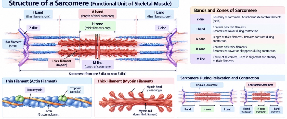
A sarcomere is the basic contractile unit of skeletal muscle. It extends from one Z disc to the next Z disc along a myofibril.
During contraction, thin actin filaments slide past thick myosin filaments, bringing the Z discs closer together and shortening the sarcomere.
The major structures of a sarcomere include the following.
Z disc: Marks the boundary of each sarcomere and provides attachment for thin filaments.
A band: Corresponds to the full length of the thick filaments. Its length remains essentially constant during contraction.
I band: Contains thin filaments without overlapping thick filaments. It becomes narrower during contraction.
H zone: The central region of the A band where thick filaments are not overlapped by thin filaments. It becomes narrower and may disappear during strong contraction.
M line: Lies near the centre of the sarcomere and helps organize and stabilize the thick filaments.
Sliding Filament Mechanism
Muscle contraction occurs through the sliding filament mechanism.
When a motor neuron stimulates a skeletal muscle fibre, an electrical signal travels along the sarcolemma and into the fibre through transverse tubules. This triggers the release of calcium ions from the sarcoplasmic reticulum.
Calcium binds to troponin, causing tropomyosin to move away from the myosin-binding sites on actin. Myosin heads can then bind to actin and generate force through repeated cross-bridge cycles.
ATP is required for myosin to detach from actin and continue cycling. As the filaments slide past one another, the sarcomere shortens.
Connective Tissue Coverings of Skeletal Muscle
The three connective tissue coverings have distinct locations.
These connective tissue layers are continuous with one another and contribute to transmitting force from muscle fibres towards tendons and ultimately to bones.
How the Organization Works During Movement
Consider lifting a book from a table.
First, the nervous system activates the appropriate motor units in the muscles involved in lifting. Within activated muscle fibres, calcium enables interactions between actin and myosin. Sarcomeres generate force, myofibrils transmit that force within each fibre, and the combined activity of many fibres produces tension in the muscle.
The connective tissue coverings help distribute this force, and the tendon transfers it to the bone. Movement occurs when the resulting forces act across a joint.
This illustrates why the organization of skeletal muscle is important: each structural level contributes to the coordinated action of the entire muscle.
Key Concepts for Examination
A whole skeletal muscle contains muscle fascicles, blood vessels, nerves and connective tissue.
Epimysium surrounds the whole muscle, perimysium surrounds each fascicle, and endomysium surrounds individual muscle fibres.
A skeletal muscle fibre is an elongated, multinucleated cell.
Myofibrils contain repeating sarcomeres.
A sarcomere extends from one Z disc to the next and is the basic contractile unit.
Actin forms the main thin filaments, while myosin forms the main thick filaments.
Calcium and ATP are essential for normal skeletal muscle contraction.
During contraction, the filaments slide past one another rather than shortening themselves.
In summary, skeletal muscle has a hierarchical organization extending from the whole muscle to fascicles, muscle fibres, myofibrils and sarcomeres. This arrangement enables the coordinated generation and transmission of force required for movement, posture and joint stability.